
Bone loss represents one of the most significant challenges facing patients considering dental implants, yet it doesn't automatically disqualify you from treatment. Modern dentistry has evolved dramatically, offering multiple pathways to successful implant placement even when significant bone deterioration has occurred. Understanding the relationship between dental implants bone loss and treatment success empowers patients to make informed decisions about their oral health and explore viable restoration options. Whether bone loss developed from periodontal disease, tooth extraction, or systemic conditions, advanced techniques now make implant dentistry accessible to more patients than ever before.
Understanding Dental Implants Bone Loss
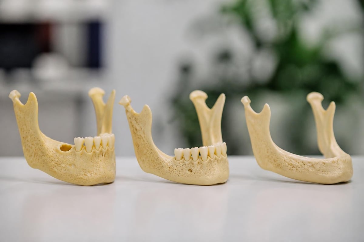
Bone loss in the jaw occurs through a natural process called resorption, where bone tissue breaks down faster than the body rebuilds it. When teeth are lost, the jawbone no longer receives stimulation from chewing forces, triggering progressive deterioration that can compromise implant placement.
The severity of dental implants bone loss varies considerably among patients. Some experience minimal reduction in bone volume, while others face substantial atrophy that alters facial structure and complicates restorative treatment. Several factors influence the rate and extent of bone deterioration, including the duration since tooth loss, age, genetics, systemic health conditions, and oral hygiene practices.
Biological Mechanisms of Bone Resorption
The jawbone maintains its density through constant mechanical stimulation. When natural teeth transmit chewing forces to the bone, specialized cells called osteoblasts continuously rebuild bone tissue. Without this stimulation, osteoclast activity exceeds osteoblast function, resulting in net bone loss.
Research indicates that bone loss accelerates during the first year following tooth extraction, with patients losing approximately 25% of bone width within the initial twelve months. This progressive deterioration continues throughout life, though at a slower rate after the first year.
Key factors accelerating bone resorption include:
- Extended periods without tooth replacement
- Periodontal disease affecting bone-supporting structures
- Denture wearing that applies uneven pressure
- Systemic conditions like osteoporosis
- Smoking and tobacco use
- Insufficient calcium and vitamin D intake
Evaluating Bone Quality and Quantity
Successful implant placement requires adequate bone volume and density to support the titanium post during osseointegration and throughout its functional lifespan. Dental professionals assess bone conditions through comprehensive imaging and clinical examination before recommending treatment approaches.
Diagnostic Imaging Technologies
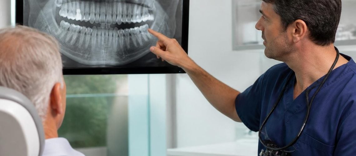
Modern diagnostic tools provide detailed three-dimensional visualization of bone architecture, enabling precise treatment planning. Cone beam computed tomography (CBCT) has become the gold standard for evaluating bone structure and density before implant placement, offering superior diagnostic accuracy compared to traditional two-dimensional radiographs.
| Imaging Method | Advantages | Limitations | Best Use Case |
|---|---|---|---|
| CBCT Scans | 3D visualization, precise measurements, radiation efficient | Higher cost than 2D imaging | Complex cases, multiple implants, bone grafting planning |
| Periapical Radiographs | Low cost, readily available, baseline documentation | Limited depth perception | Follow-up monitoring, single-tooth evaluation |
| Panoramic X-rays | Full jaw overview, screening tool | Distortion, less detailed | Initial assessment, general screening |
The comprehensive imaging recommendations established by professional organizations emphasize obtaining baseline intraoral radiographs immediately after implant placement and periodic follow-up imaging to monitor bone levels around implants.
Bone Density Classification
Dental professionals classify bone density using a standardized system that guides treatment planning. Dense cortical bone provides excellent initial stability but requires careful surgical technique, while softer trabecular bone may necessitate modified protocols or augmentation procedures.
Understanding your bone classification helps determine whether immediate implant placement is feasible or if preparatory procedures like grafting are necessary. At Dental Plus Clinic, comprehensive evaluations ensure personalized treatment plans that address individual bone conditions.
Risk Factors Contributing to Dental Implants Bone Loss
Multiple biological, mechanical, and behavioral factors influence bone health around dental implants. Identifying these risks before treatment enables proactive management strategies that improve long-term outcomes.
Biological Risk Factors
Systemic health conditions significantly impact bone metabolism and healing capacity. Research examining osteoporosis and anti-resorptive medications demonstrates that patients with compromised bone metabolism face elevated risks for marginal bone loss around implants, though success rates remain high with appropriate modifications.
Significant biological contributors include:
- Periodontal disease history – Prior gum disease indicates susceptibility to bacterial infections that can affect implants
- Diabetes mellitus – Elevated blood glucose impairs healing and increases infection risk
- Autoimmune conditions – Disorders affecting immune function may compromise osseointegration
- Medication effects – Bisphosphonates, immunosuppressants, and certain other drugs influence bone metabolism
- Nutritional deficiencies – Inadequate calcium, vitamin D, and protein intake undermines bone health
Mechanical and Prosthetic Considerations
The design and fabrication of implant restorations directly influence stress distribution and bone preservation. A comprehensive review of prosthetic design factors highlights how emergence profiles, contour accuracy, and component selection affect peri-implant health and marginal bone stability.
Excessive forces from poorly balanced restorations, inadequate crown contours that trap bacteria, and cement residue left during crown placement all contribute to progressive bone loss. Modern digital design protocols minimize these risks through precise engineering and careful material selection.
Prevention Strategies for Maintaining Bone Health
Proactive measures before, during, and after implant placement significantly reduce the risk of dental implants bone loss. Evidence-based protocols established through consensus recommendations on peri-implant disease prevention guide clinical practice and patient education.
Pre-Surgical Optimization
Preparing the patient's oral and systemic health before surgery creates optimal conditions for implant success. This preparation phase addresses modifiable risk factors and establishes baseline health parameters.
Essential pre-surgical steps include:
- Comprehensive periodontal therapy to eliminate active gum disease
- Smoking cessation programs for tobacco users
- Blood glucose optimization for diabetic patients
- Nutritional counseling to ensure adequate protein and micronutrient intake
- Bone density assessment for patients with osteoporosis risk
Patients can explore affordable preventive care options that support optimal oral health before implant treatment.
Surgical Protocol Refinements
Advanced surgical techniques minimize trauma to existing bone and optimize initial implant stability. Careful site preparation, atraumatic extraction protocols when removing hopeless teeth, and immediate implant placement in appropriate cases all contribute to bone preservation.
The surgical approach selected depends on individual bone anatomy, implant position requirements, and restoration goals. Experienced clinicians adapt techniques based on real-time observations during surgery, making adjustments that protect bone integrity.
Bone Augmentation Solutions
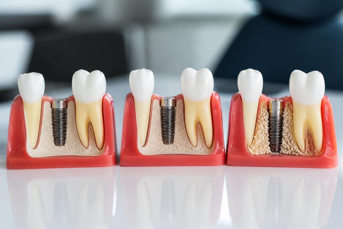
When existing bone volume proves insufficient for standard implant placement, regenerative procedures can rebuild lost bone tissue. These techniques have transformed implant dentistry, enabling treatment for patients previously considered unsuitable candidates.
Bone Grafting Procedures
Bone grafting involves placing bone material at deficient sites to stimulate new bone formation. The grafting material serves as a scaffold that guides the patient's natural bone cells to regenerate tissue, gradually replacing the graft material with living bone.
Several graft material sources exist, each with distinct advantages:
| Graft Type | Source | Advantages | Considerations |
|---|---|---|---|
| Autograft | Patient's own bone | Best biocompatibility, no rejection risk | Requires second surgical site |
| Allograft | Processed human donor bone | No harvest site needed, readily available | Slightly lower incorporation rate |
| Xenograft | Bovine or porcine bone | Abundant supply, cost-effective | Slower remodeling, some cultural concerns |
| Alloplast | Synthetic materials | No disease transmission risk, unlimited supply | Variable long-term performance |
Sinus Lift Procedures
The maxillary sinuses often limit implant placement in the upper back jaw regions. Sinus augmentation elevates the sinus membrane and places bone graft material beneath it, creating adequate bone height for implant support.
Two approaches exist for sinus augmentation. The lateral window technique provides access for substantial bone volume addition, while the osteotome approach offers a less invasive option for cases requiring moderate augmentation.
Ridge Expansion Techniques
Narrow bone ridges can be widened through controlled expansion procedures that gradually separate the bone plates. This approach preserves existing bone while creating space for implant placement, often allowing simultaneous implant insertion.
Alternative Implant Solutions for Severe Bone Loss
Patients with extensive bone atrophy may benefit from specialized implant designs that work within available bone anatomy. These alternatives offer viable pathways to stable tooth replacement without extensive grafting procedures.
Zygomatic Implants
Zygomatic implants anchor in the cheekbone (zygoma) rather than the maxillary bone, bypassing severely atrophied upper jaw regions. This approach enables immediate full-arch restoration for patients who would otherwise require extensive bone grafting with prolonged treatment timelines.
The technique demands specialized training and precise surgical execution, but it provides life-changing results for patients with significant maxillary bone loss. Success rates exceed 95% in experienced hands, with patients enjoying stable, functional restorations.
Subperiosteal Implants
Recent advances in additively manufactured subperiosteal implants have renewed interest in this approach for patients with severe bone atrophy. Custom-designed frameworks sit beneath the gum tissue but above the bone, distributing forces across remaining bone architecture.
Digital scanning and 3D printing technologies enable precise fabrication of patient-specific subperiosteal devices. While less common than endosteal implants, they offer solutions when bone reconstruction proves impractical or medically contraindicated.
All-On-4 and All-On-6 Treatment Concepts
Full-arch implant protocols strategically position four or six implants to support complete dental arches, often utilizing posterior implants at angles that engage more dense bone regions. This approach maximizes available bone volume while minimizing the need for grafting procedures.
Patients interested in comparing these options can review detailed information about All-On-4 versus All-On-6 dental implants to understand which configuration best suits their needs.
Managing Existing Dental Implants Bone Loss
Even successfully integrated implants may experience progressive bone loss years after placement. Early detection and intervention preserve implant function and prevent more extensive bone deterioration.
Peri-Implantitis Recognition and Treatment
Peri-implantitis represents inflammatory disease affecting tissues around dental implants, characterized by progressive bone loss and potential implant failure. The condition shares similarities with periodontitis but progresses more rapidly due to implants' lack of periodontal ligament protection.
Warning signs of peri-implantitis include:
- Bleeding when cleaning around implants
- Gum recession exposing implant threads
- Pus discharge from peri-implant tissues
- Increasing tooth mobility
- Discomfort or pain around implant sites
Early-stage peri-implant disease (peri-implant mucositis) responds well to non-surgical therapy involving professional cleaning and improved home care. Advanced cases with significant bone loss may require surgical intervention approaches including regenerative procedures or resective techniques to re-establish healthy bone architecture.
Non-Surgical Maintenance Protocols
Regular professional maintenance appointments enable early detection of problems before extensive bone loss develops. During these visits, dental professionals assess bone levels through radiographic evaluation, probe pocket depths around implants, and remove bacterial deposits from implant surfaces.
Effective home care practices include:
- Daily cleaning around implant restorations using soft-bristled brushes
- Interdental cleaning with specialized implant floss or brushes
- Antimicrobial mouth rinses when recommended by dental professionals
- Avoiding abrasive cleaning products that damage implant surfaces
- Monitoring for early signs of inflammation or discomfort
Surgical Regeneration Options
When dental implants bone loss advances despite non-surgical therapy, surgical intervention may preserve the implant and restore lost bone. Regenerative procedures apply similar principles to those used during initial implant placement, utilizing bone grafts and barrier membranes to encourage new bone formation.
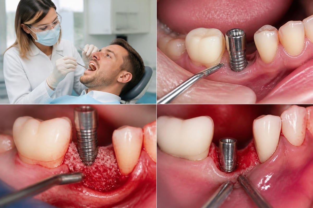
Success rates for regenerative treatment of peri-implantitis vary based on defect configuration, patient compliance with maintenance protocols, and elimination of causative factors. Realistic expectations and commitment to ongoing care significantly influence outcomes.
Systemic Health Optimization for Implant Success
Overall health status profoundly impacts bone metabolism, healing capacity, and long-term implant survival. Addressing systemic conditions before and after implant placement reduces complications and supports bone preservation.
Diabetes Management
Patients with diabetes face elevated risks for infection and delayed healing, both of which can compromise implant outcomes. However, well-controlled diabetes does not preclude successful implant treatment. Maintaining hemoglobin A1c levels below 7% significantly improves healing and reduces peri-implant disease risk.
Coordination between dental and medical providers ensures optimal glycemic control during the surgical and healing phases. Some patients benefit from temporary medication adjustments or more frequent monitoring during implant treatment.
Osteoporosis Considerations
Osteoporosis affects bone density throughout the body, including the jaw. While this condition influences bone quality, it does not prevent successful implant placement. Understanding the impact of osteoporosis and anti-resorptive medications on implant outcomes helps guide treatment timing and protocol modifications.
Patients taking bisphosphonates or other anti-resorptive medications require individualized assessment. The duration of medication use, administration route (oral versus intravenous), and specific drug properties all influence surgical planning and healing expectations.
Nutritional Support for Bone Health
Adequate nutrition provides essential building blocks for bone formation and repair. Protein intake supports tissue healing, while calcium and vitamin D optimize bone mineralization. Additional nutrients including vitamin C, vitamin K, magnesium, and phosphorus contribute to bone health.
Nutritional recommendations for implant patients:
- Daily protein intake of 1.0-1.2 grams per kilogram body weight
- Calcium supplementation to achieve 1000-1200 mg daily total intake
- Vitamin D levels maintained above 30 ng/mL through supplementation
- Adequate hydration supporting nutrient transport and waste removal
- Limitation of substances impairing bone metabolism (excessive caffeine, alcohol)
Long-Term Success Factors and Maintenance
Dental implants bone loss prevention requires lifelong commitment to oral hygiene, professional maintenance, and risk factor management. Recent research summarizing risk factors and prevention strategies emphasizes that biological, mechanical, and prosthetic factors all contribute to long-term bone stability around implants.
Professional Maintenance Schedules
Regular professional care enables early intervention when problems develop, preventing minor issues from progressing to significant bone loss. Most implant patients benefit from professional maintenance appointments every three to six months, with intervals adjusted based on individual risk factors.
During maintenance visits, dental professionals perform the following evaluations:
| Assessment Component | Purpose | Frequency |
|---|---|---|
| Radiographic imaging | Monitor bone levels | Annually or as indicated |
| Probing depth measurement | Detect early inflammation | Each maintenance visit |
| Plaque and calculus removal | Eliminate bacterial irritants | Every 3-6 months |
| Occlusion evaluation | Identify excessive forces | Annually or when symptoms arise |
| Home care review | Optimize patient techniques | Each maintenance visit |
Restoration Longevity and Replacement
Implant restorations have finite lifespans, with crowns and bridges typically requiring replacement every 10-15 years due to normal wear. Timely restoration replacement prevents complications like fractured components or cement loss that can contribute to bone loss.
Patients exploring the financial aspects of implant treatment can learn about why dental implants represent a long-term investment in oral health and quality of life.
Technological Advances Improving Outcomes
Modern dentistry continuously evolves, with new technologies and techniques enhancing implant success rates even in challenging bone situations. Digital workflows, improved surface treatments, and biomimetic materials contribute to better outcomes for patients facing bone deficiency.
Digital Planning and Guided Surgery
Computer-aided design and manufacturing (CAD/CAM) technologies enable virtual implant placement before surgery. Clinicians evaluate bone volume, plan optimal implant positions, and identify anatomical structures requiring protection. This digital workflow translates to surgical guides that direct precise implant placement according to the virtual plan.
Guided surgery reduces surgical time, minimizes tissue trauma, and improves accuracy in complex cases. Patients benefit from more predictable outcomes and often faster recovery periods.
Surface Technology Innovations
Implant surface modifications enhance bone cell attachment and accelerate osseointegration. Modern surface treatments create micro- and nano-scale topographies that promote protein adsorption and cellular activity, improving integration in compromised bone quality.
Hydrophilic surfaces demonstrate particular advantages in challenging situations, attracting bone cells more effectively than traditional surfaces. These innovations expand treatment possibilities for patients with less-than-ideal bone conditions.
Biomaterial Development
Advanced bone graft materials and barrier membranes incorporate growth factors and bioactive molecules that stimulate bone formation. These enhanced materials accelerate healing and improve regenerative outcomes compared to traditional grafting approaches.
Platelet-rich fibrin (PRF) and platelet-rich plasma (PRP) concentrate the patient's own growth factors, providing biological enhancement for bone grafting procedures. While research continues evaluating long-term benefits, many clinicians report improved healing and reduced complications when incorporating these technologies.
Financial Considerations and Treatment Planning
Cost represents a significant consideration for patients evaluating implant treatment, particularly when bone grafting or other preparatory procedures add to overall investment. Understanding treatment costs and available financial options helps patients make informed decisions.
Treatment Cost Components
Implant treatment involves multiple phases, each with associated costs. Diagnostic imaging, surgical procedures, bone grafting when necessary, implant placement, healing components, and final restorations all contribute to total investment.
Typical cost breakdown includes:
- Initial consultation and diagnostic imaging
- Bone grafting procedures (when required)
- Implant surgical placement
- Healing abutments and temporary restorations
- Final crown or bridge fabrication
- Follow-up care and maintenance
Patients can explore options for dental implants without insurance to understand available payment plans and financing alternatives.
Value Assessment
While implant treatment requires substantial upfront investment, the long-term value often exceeds alternative treatments. Properly maintained implants can last decades, avoiding the recurring replacement costs associated with conventional bridges and removable dentures.
Additional value considerations include improved chewing function, better nutrition from dietary flexibility, enhanced confidence from stable teeth, and preservation of remaining natural teeth that might otherwise support bridges.
Patient Selection and Treatment Planning
Not every patient with bone loss represents an ideal implant candidate, and honest assessment of success probability guides ethical treatment recommendations. Comprehensive evaluation considers medical history, anatomical limitations, behavioral factors, and patient expectations.
Candidacy Assessment
Ideal implant candidates demonstrate good general health, adequate bone volume, healthy gum tissues, commitment to oral hygiene, and realistic expectations. Patients with controlled systemic diseases, moderate bone loss amenable to grafting, or lifestyle factors like smoking may still achieve success but require modified protocols and enhanced monitoring.
Absolute contraindications to implant treatment include:
- Active cancer treatment affecting bone metabolism
- Recent high-dose radiation to jaw regions
- Uncontrolled diabetes with poor healing capacity
- Severe bone loss insufficient for implant support without extensive reconstruction
- Inability to comply with oral hygiene requirements
Realistic Expectation Setting
Successful implant treatment requires alignment between patient expectations and achievable outcomes. Comprehensive consultation addresses treatment duration, number of procedures required, healing periods, temporary tooth replacement options, and long-term maintenance commitments.
Patients should understand that bone grafting extends treatment timelines, often adding four to six months for graft maturation before implant placement. While this delay may seem frustrating, it creates the foundation for long-term implant success.
Comparing Alternative Tooth Replacement Options
When extensive bone loss complicates implant treatment or patient factors suggest alternative approaches, other restoration options merit consideration. Each approach offers distinct advantages and limitations based on individual circumstances.
Fixed Bridges
Conventional bridges replace missing teeth by crowning adjacent natural teeth and suspending artificial teeth between the abutments. This approach avoids surgical procedures and provides fixed, non-removable restorations.
However, bridges require permanent alteration of healthy adjacent teeth, concentrate chewing forces on supporting teeth potentially causing accelerated wear, and fail to prevent continued bone loss in edentulous areas. Average bridge lifespan ranges from 10-15 years before replacement becomes necessary.
Patients can review comparisons of dental implants versus veneers for missing teeth to understand different restoration approaches.
Removable Dentures
Complete and partial dentures provide economical tooth replacement without surgery. Modern dentures offer improved aesthetics and function compared to previous generations, with various material and design options available.
Denture limitations include reduced chewing efficiency (approximately 25% of natural tooth function), potential discomfort or movement during eating and speaking, accelerated bone loss from pressure on soft tissues, and frequent need for relining or replacement as bone continues resorbing.
Implant-Supported Overdentures
Hybrid approaches combine implant stability with removable prosthetics, offering a middle ground between conventional dentures and fixed implant restorations. Two to four strategically placed implants retain removable dentures, dramatically improving stability and function while requiring fewer implants than fixed restorations.
This option suits patients with extensive bone loss who want improved function and stability without extensive grafting procedures. The removable design facilitates cleaning and allows economical component replacement when needed.
Understanding dental implants bone loss opens pathways to successful tooth replacement even when bone deficiency seems discouraging. Modern diagnostic tools, advanced surgical techniques, and evidence-based maintenance protocols enable most patients to achieve stable, functional implant restorations that preserve remaining bone and restore confident smiles. Whether you're exploring implant treatment for the first time or managing existing restorations, Dental Plus Clinic provides comprehensive evaluation, personalized treatment planning, and compassionate care across our five Texas locations in Beeville, Seguin, New Braunfels, Leander, and Converse to help you achieve optimal oral health outcomes.