
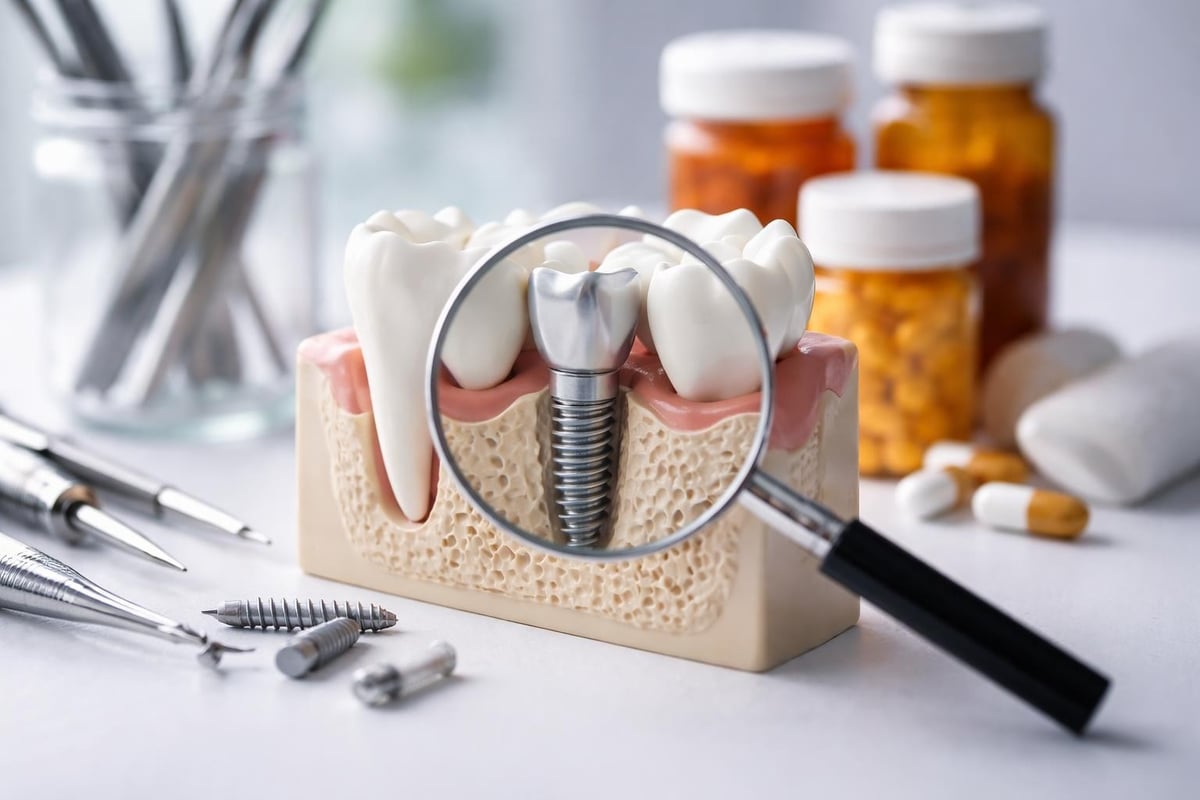
Dental implants have revolutionized modern dentistry, offering patients a permanent solution for missing teeth that looks, feels, and functions like natural teeth. With success rates exceeding 95%, implants have become the gold standard for tooth replacement. However, like any surgical procedure, dental implants carry potential risks that patients should understand before committing to treatment. While complications are relatively rare when performed by experienced professionals, being informed about the risks and complications of dental implants empowers patients to make educated decisions about their oral health and recognize warning signs early if problems arise.
Understanding Common Infection Risks
Infection represents one of the most significant concerns following implant placement. The surgical nature of dental implant procedures creates an opening in the gum tissue and bone, providing a potential pathway for bacteria to enter.
Immediate Post-Surgical Infections
Infections occurring within days or weeks after surgery typically result from contamination during the procedure or inadequate post-operative care. Symptoms include swelling, persistent pain beyond normal healing discomfort, pus discharge, and fever. These early infections require prompt antibiotic treatment and, in severe cases, temporary implant removal.
Risk factors for post-surgical infections include:
- Poor oral hygiene before and after surgery
- Compromised immune system function
- Uncontrolled diabetes
- Smoking and tobacco use
- Pre-existing periodontal disease
Modern surgical protocols significantly reduce infection rates. Sterile techniques, prophylactic antibiotics, and advanced imaging for precise placement all contribute to safer procedures. At facilities prioritizing patient safety, infection rates remain exceptionally low when patients follow post-operative instructions carefully.
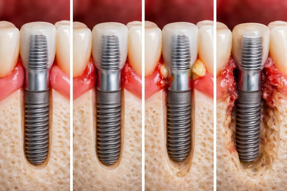
Peri-Implantitis: The Long-Term Threat
Peri-implantitis represents a progressive inflammatory condition affecting the soft and hard tissues surrounding dental implants. Unlike early infections, peri-implantitis develops months or years after successful integration, making it particularly insidious.
This condition begins with peri-mucositis, inflammation of the soft tissue around the implant without bone loss. If untreated, it progresses to peri-implantitis, characterized by bone deterioration that threatens implant stability. The condition affects an estimated 10-20% of implants over a 10-year period.
| Peri-Mucositis | Peri-Implantitis |
|---|---|
| Soft tissue inflammation only | Inflammation plus bone loss |
| Reversible with treatment | May require surgical intervention |
| No bleeding on probing depth | Increased probing depths with bleeding |
| Implant remains stable | Progressive implant loosening |
Prevention requires meticulous oral hygiene, regular professional cleanings, and monitoring by dental professionals. Patients who maintain consistent dental check-ups detect early warning signs before significant damage occurs.
Nerve Damage and Sensory Complications
Nerve injury constitutes another serious concern among the risks and complications of dental implants, particularly for lower jaw placements. The inferior alveolar nerve runs through the lower jaw, providing sensation to the lower lip, chin, and teeth on that side.
Types of Nerve Injuries
Nerve damage ranges from temporary numbness to permanent sensory loss. Neuropraxia, the mildest form, involves temporary nerve compression causing numbness that typically resolves within weeks to months. More severe injuries include axonotmesis, where nerve fibers are damaged but the nerve sheath remains intact, and neurotmesis, complete nerve severance requiring surgical intervention.
According to Medical News Today, nerve damage symptoms include:
- Persistent numbness or tingling in the lip, chin, or tongue
- Altered taste perception
- Difficulty speaking or eating
- Pain or burning sensations in affected areas
- Drooling due to reduced lip sensation
Advanced diagnostic imaging, particularly cone beam computed tomography (CTCT), allows precise mapping of nerve locations before surgery. Experienced surgeons maintain safe distances from vital structures, significantly reducing nerve injury risk. When choosing between materials like zirconia versus titanium implants, proper placement matters more than material selection for avoiding nerve complications.
Prevention Through Careful Planning
Three-dimensional imaging technology has revolutionized implant planning. Surgeons can now visualize exact nerve positions, bone density, and optimal implant angles before making the first incision. Digital planning software even creates surgical guides ensuring implants are placed precisely as planned.
Patient selection also plays a crucial role. Individuals with limited bone height in areas near nerves may require bone grafting procedures to create adequate space between the implant and nerve structures. This additional step, while extending treatment time, substantially reduces nerve damage risk.
Implant Failure and Integration Problems
Implant failure, though uncommon, represents perhaps the most disappointing complication patients face. Failure occurs in two distinct phases: early failure before osseointegration completes, and late failure after years of successful function.
Early Integration Failures
Early failures typically happen within the first few months after placement, during the critical osseointegration period when bone cells grow onto the implant surface. Multiple factors can disrupt this delicate process.
Common causes of early implant failure:
- Insufficient bone quantity or quality
- Excessive loading before complete integration
- Implant contamination during placement
- Poor primary stability at insertion
- Patient smoking or tobacco use
- Uncontrolled systemic diseases
Movement during healing proves particularly detrimental. Even microscopic motion prevents proper bone formation around the implant. This explains why temporary restorations must avoid placing pressure on healing implants, and why patients receive strict dietary restrictions during recovery.
Bone quality significantly influences success rates. The jawbone is classified into four density types, with Type I being densest and Type IV least dense. Lower density bone provides less initial stability and requires longer healing periods. Understanding your bone quality helps set realistic expectations for treatment timelines.
Late-Stage Implant Failures
Late failures occur after successful integration, sometimes years after placement. These failures often result from biological or mechanical factors rather than surgical issues.
Biological late failures frequently stem from peri-implantitis, discussed earlier. The gradual bone loss eventually compromises implant support, leading to mobility and failure. Mechanical failures include implant fracture, abutment loosening, or prosthetic component failure.
| Early Failure Factors | Late Failure Factors |
|---|---|
| Poor surgical technique | Peri-implantitis development |
| Inadequate bone quality | Occlusal overload |
| Premature loading | Implant fracture |
| Infection during healing | Component loosening |
| Patient smoking | Poor oral hygiene maintenance |
The latest dental implant technology incorporates features designed to resist both early and late failures. Surface treatments enhance osseointegration, while improved materials resist fracture under normal chewing forces.
Sinus-Related Complications
Upper jaw implants pose unique risks related to the maxillary sinuses, air-filled spaces located above the upper back teeth. Insufficient bone between the sinus floor and the oral cavity can lead to several complications.
Sinus Membrane Perforation
During upper implant placement or sinus lift procedures, the delicate Schneiderian membrane lining the sinus can tear. Small perforations often heal without intervention, but larger tears may cause chronic sinusitis, sinus infections, or implant migration into the sinus cavity.
Symptoms of sinus complications include:
- Nasal congestion or discharge
- Facial pressure or pain
- Headaches localized to the sinus area
- Altered sense of smell
- Metallic taste in the mouth
Prevention requires thorough pre-operative assessment using 3D imaging to measure bone height accurately. When bone height proves insufficient, sinus augmentation procedures create adequate space for safe implant placement. While adding treatment complexity, these preventive measures virtually eliminate sinus perforation risks.
Chronic Sinus Issues
Even without perforation, implants placed too close to the sinus floor can trigger chronic inflammation. The proximity alters normal sinus drainage patterns, potentially leading to recurring infections. This complication emphasizes the importance of maintaining appropriate safety margins during placement.
Experienced clinicians account for anatomical variations between patients. Some individuals naturally have larger sinuses or thinner bone, requiring modified approaches. Customized treatment planning addresses these individual factors, reducing complication rates substantially.
Bone Loss and Gum Recession
Progressive bone loss around dental implants can occur even without infection. This phenomenon, distinct from peri-implantitis, results from biomechanical factors and stress distribution.
Stress Shielding and Bone Remodeling
Bone responds to functional stress through constant remodeling. When stress distribution around an implant differs significantly from natural tooth support, bone may resorb in areas receiving insufficient stimulation. This “stress shielding” effect depends on implant design, placement angle, and prosthetic loading.
Proper implant positioning ensures forces distribute naturally through surrounding bone. Implants angled incorrectly or subjected to excessive lateral forces experience accelerated bone loss. This explains why prosthodontic planning matters as much as surgical placement.
Factors contributing to bone loss around implants:
- Excessive occlusal forces from grinding or clenching
- Poorly designed prosthetics creating leverage
- Inadequate implant diameter for the restoration size
- Systemic conditions affecting bone metabolism
- Lack of functional stimulation in adjacent areas
The comparison between All-on-4 and All-on-6 approaches demonstrates how implant number and positioning affect long-term bone preservation. Additional implants often distribute forces more evenly, potentially reducing stress-related bone loss.
Gum Tissue Recession
Soft tissue recession exposes implant components, creating aesthetic concerns and increasing infection risk. Recession occurs more commonly with thin tissue biotypes, inadequate attached gingiva, or poor oral hygiene allowing plaque accumulation.
| Risk Factor | Impact on Recession |
|---|---|
| Thin gum tissue | Higher recession susceptibility |
| Lack of keratinized tissue | Reduced tissue stability |
| Aggressive brushing | Mechanical tissue trauma |
| Prominent implant positioning | Tissue tension and thinning |
| Smoking | Impaired tissue healing and health |
Prevention strategies include soft tissue grafting at implant placement to increase tissue thickness, proper implant positioning to support overlying tissue, and patient education on gentle hygiene techniques. When recession occurs, additional grafting procedures can restore coverage, though success depends on the extent of tissue loss.
Patient-Specific Risk Factors
Individual patient characteristics significantly influence the risks and complications of dental implants. Understanding personal risk factors allows for tailored treatment approaches that maximize success probability.
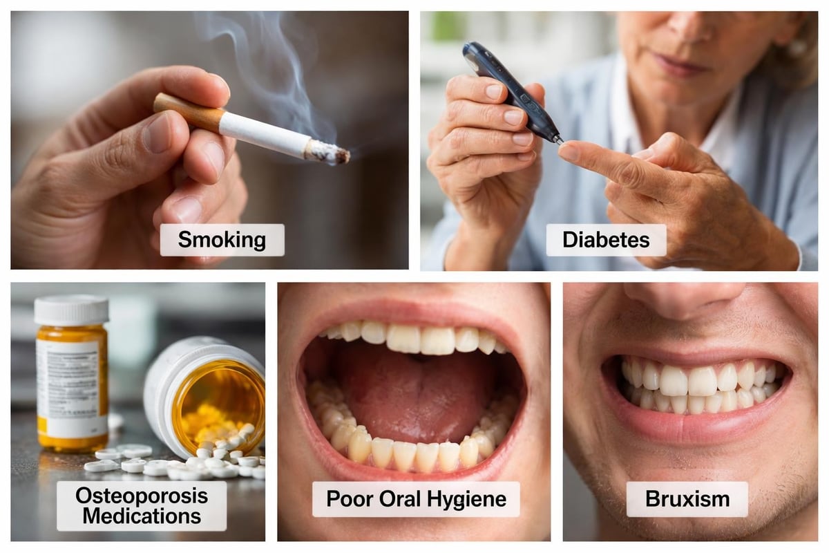
Medical Conditions Affecting Outcomes
Certain systemic diseases complicate implant healing and long-term success. Diabetes, particularly when poorly controlled, impairs wound healing and increases infection susceptibility. Patients with HbA1c levels above 7% face substantially higher complication rates than those with well-managed blood sugar.
Osteoporosis and medications treating it present complex considerations. While osteoporosis itself doesn’t necessarily contraindicate implants, bisphosphonate medications used to treat the condition increase risk of osteonecrosis, a serious bone death condition. The risk varies with medication type, dosage, and duration of use.
Autoimmune disorders affecting connective tissue, such as rheumatoid arthritis or lupus, may impair healing and integration. Immunosuppressant medications further complicate matters by reducing the body’s ability to fight infection and heal tissue.
Lifestyle Factors and Behavioral Risks
According to research highlighted by Oceanic Dental, smoking ranks among the most significant modifiable risk factors for implant failure. Nicotine constricts blood vessels, reducing oxygen and nutrient delivery to healing tissues. Carbon monoxide further impairs healing by displacing oxygen in the bloodstream.
Statistics demonstrate the impact clearly:
- Non-smokers: 95-98% success rate
- Light smokers (less than 10 cigarettes daily): 88-92% success rate
- Heavy smokers (more than 10 cigarettes daily): 80-85% success rate
Smoking cessation, even temporarily around surgery, dramatically improves outcomes. Most clinicians recommend stopping at least two weeks before surgery and avoiding tobacco throughout the healing period.
Bruxism, chronic teeth grinding and clenching, subjects implants to excessive forces. While natural teeth have periodontal ligaments that cushion impact, implants connect directly to bone. This rigid connection makes them more vulnerable to stress fractures and accelerated bone loss. Night guards and stress management help protect implants in bruxism patients.
Prosthetic and Mechanical Complications
Beyond biological issues, mechanical problems with implant components can compromise function and aesthetics. These complications range from minor annoyances to situations requiring complete implant replacement.
Abutment and Screw Loosening
The abutment connects the implant fixture to the final crown or bridge. This connection relies on precise torque specifications and proper component fit. Over time, chewing forces can loosen these connections, causing clicking sounds, movement, or complete separation.
Screw loosening occurs more frequently with single-unit restorations than with multi-unit bridges, as bridges distribute forces across multiple implants. External hex connections show higher loosening rates than internal connections, which provide better mechanical stability.
Signs of abutment loosening include:
- Clicking or movement when chewing
- Food packing around the restoration
- Changes in how teeth fit together
- Visible gap between crown and gum line
- Discomfort when biting
Regular professional maintenance allows early detection and simple retorquing before complications develop. Most loosening incidents occur within the first year after placement as components settle under functional loads.
Prosthetic Material Failures
Crown or bridge materials occasionally chip, crack, or fracture. Porcelain, while aesthetic, proves more brittle than natural tooth enamel. Excessive forces, particularly in bruxism patients, can cause porcelain fracture exposing underlying metal frameworks.
Acrylic materials used in temporary restorations or some full-arch solutions may wear or stain over time. While durable enough for function, they require eventual replacement to maintain aesthetics and proper bite relationships.
Material selection balances aesthetics, strength, and cost. The choice between cheap and premium dental implants often comes down to prosthetic components as much as implant fixtures. Premium materials and precise manufacturing reduce mechanical complication rates.
Complete Implant Fracture
Though rare, implant fixtures can fracture under extreme or prolonged excessive loading. Fracture occurs more commonly with narrow-diameter implants, particularly in posterior regions experiencing highest chewing forces. Once fractured, implants require complete removal and potential bone grafting before replacement.
| Implant Diameter | Fracture Risk | Typical Applications |
|---|---|---|
| Narrow (3.0-3.5mm) | Higher risk | Anterior teeth, limited spaces |
| Standard (3.75-4.5mm) | Low risk | Most single-tooth replacements |
| Wide (5.0-6.0mm) | Very low risk | Molars, multi-unit restorations |
Proper implant sizing for the restoration site and expected forces represents critical planning. Placing narrow implants in molar positions to avoid bone grafting may save initial costs but increases long-term fracture risk substantially.
Aesthetic Complications and Soft Tissue Issues
Beyond function, implants must blend naturally with surrounding teeth and gums. Aesthetic complications, while not affecting health directly, significantly impact patient satisfaction.
Gingival Discoloration
Titanium implants placed too close to thin gum tissue can create a grayish discoloration visible through the tissue. This “graying” effect particularly concerns anterior restorations where aesthetics matter most. The phenomenon results from metal showing through translucent tissue.
Prevention strategies include:
- Maintaining adequate tissue thickness through grafting
- Proper implant positioning slightly deeper and more palatal
- Using zirconia abutments instead of titanium in aesthetic zones
- Selecting appropriate restoration materials that block underlying color
Once graying occurs, treatment options prove limited. Tissue grafting to increase thickness may help, but results vary. This complication underscores the importance of aesthetic planning from the beginning rather than attempting corrections later.
Papilla Loss and Black Triangles
The triangular gum tissue between teeth, called papillae, fills spaces naturally. Implants lack the biological support structures that maintain papillae around natural teeth. The distance from the contact point between teeth to underlying bone determines whether papillae fill completely.
When this distance exceeds approximately 5mm, papillae rarely regenerate completely, leaving dark spaces. These “black triangles” create aesthetic concerns and food trapping. Prevention requires proper implant placement and maintaining adequate bone levels between adjacent teeth.
Restoration Emergence Profile Issues
The way a crown emerges from gum tissue affects both aesthetics and cleanliness. Overly bulbous crowns create plaque traps and unnatural appearance. Conversely, under-contoured restorations leave gaps where food accumulates and tissue appears unsupported.
Achieving optimal emergence requires skilled prosthetic work. Custom abutments shaped specifically for each site provide better tissue support than stock components. Digital design and milling allow precise customization improving both function and appearance.
Rare But Serious Complications
While most risks and complications of dental implants involve relatively minor issues, certain rare complications demand awareness despite low occurrence rates.
Implant Migration
Extremely rarely, implants can migrate from their placement site. Upper jaw implants may shift into the sinus cavity if bone integrity becomes compromised. Migration typically indicates severe bone loss or initial placement in inadequate bone.
Symptoms vary depending on migration location but may include sudden position changes, altered sensation, or sinus symptoms. Treatment requires implant removal and bone reconstruction before considering replacement.
Hemorrhage and Vascular Injury
As noted by experts discussing surgical risks, excessive bleeding during or after surgery, though uncommon, can occur if blood vessels are damaged. The floor of the mouth contains significant blood vessels that, if perforated, can cause substantial hemorrhage.
Careful surgical planning and awareness of anatomical structures prevent most vascular injuries. Immediate recognition and appropriate intervention control bleeding when it occurs. Patients taking blood thinners face elevated bleeding risks and require special protocols coordinated with their physicians.
Allergic Reactions to Implant Materials
True titanium allergy remains exceptionally rare, affecting less than 1% of the population. However, some individuals experience hypersensitivity reactions causing chronic inflammation around implants without infection. Symptoms include persistent swelling, discomfort, and implant mobility despite proper placement.
Pre-operative allergy testing is not routine due to low prevalence, but patients with known metal sensitivities should discuss concerns with their surgeon. Zirconia implants offer an alternative for patients with confirmed titanium sensitivity.
Minimizing Risks Through Proper Patient Selection and Planning
Understanding the risks and complications of dental implants enables both patients and clinicians to implement preventive strategies. Comprehensive evaluation identifies high-risk situations requiring modified approaches or alternative treatments.
Thorough Pre-Operative Assessment
Complete medical and dental histories reveal risk factors requiring management. Blood tests may assess diabetes control, while bone density scans evaluate osteoporosis. Periodontal evaluation ensures gum disease treatment precedes implant placement.
Essential pre-operative evaluations include:
- Comprehensive medical history review
- Detailed oral examination and periodontal assessment
- 3D imaging to evaluate bone quantity and quality
- Analysis of bite relationships and opposing teeth
- Discussion of patient expectations and concerns
- Review of medications and potential interactions
Advanced imaging proves invaluable for surgical planning. Cone beam CT scans provide three-dimensional views showing exact bone dimensions, nerve locations, and sinus positions. This information allows virtual implant placement, identifying potential complications before surgery begins.
Risk Mitigation Strategies
When risk factors exist, specific strategies reduce complication probability. Diabetic patients benefit from achieving optimal glucose control before surgery. Smokers who quit, even temporarily, experience substantially better outcomes.
Antibiotic prophylaxis prevents infection in high-risk patients. Bone grafting augments deficient sites, creating adequate volume for safe placement. Antimicrobial mouth rinses reduce bacterial loads before and after surgery.
Treatment sequencing matters significantly. Addressing periodontal disease before implant placement prevents bacterial colonization of new implants. Extracting hopeless teeth and allowing complete healing creates optimal conditions for implant integration.
The Role of Experience and Technology
Implant success correlates strongly with surgeon experience and facility technology. High-volume practitioners develop refined techniques through repetition, while advanced equipment enables precision impossible with traditional methods.
Surgeon Expertise and Training
Dental implant placement requires specialized training beyond general dentistry. Periodontists, oral surgeons, and prosthodontists receive extensive implant education during residency programs. General dentists pursuing implant placement should complete comprehensive continuing education courses.
Experience reduces complication rates through better surgical technique, improved case selection, and enhanced problem recognition. According to Cleveland Implant Institute research, practitioners placing fewer than 50 implants annually show higher complication rates than those performing several hundred procedures.
Technology Reducing Complications
Computer-guided implant surgery represents a significant advancement in safety and precision. Surgical guides, created from digital treatment plans, direct drill placement with sub-millimeter accuracy. This precision reduces nerve damage risk, ensures optimal positioning, and minimizes surgical trauma.
Digital impressions eliminate discomfort of traditional molds while providing superior accuracy for prosthetic fabrication. Better-fitting restorations reduce mechanical complications and improve long-term success.
| Traditional Approach | Technology-Enhanced Approach |
|---|---|
| Freehand surgical placement | Computer-guided surgery |
| Physical impression materials | Digital scanning |
| 2D radiographs for planning | 3D cone beam imaging |
| Stock abutment components | Custom-milled abutments |
| Manual torque estimation | Electronic torque control |
Laser therapy enhances healing and reduces infection risk through antimicrobial effects and improved tissue response. Platelet-rich fibrin derived from patient blood accelerates healing and improves bone formation around implants.
Managing Complications When They Occur
Despite best efforts, complications sometimes develop. Early recognition and appropriate intervention minimize long-term consequences and often salvage implants that might otherwise fail.
Monitoring and Early Detection
Regular professional monitoring allows early complication detection. Annual examinations should include:
- Visual assessment of soft tissues around implants
- Probing depths to detect bone loss
- Radiographs evaluating bone levels
- Stability testing to identify mobility
- Bite analysis checking for excessive forces
Patients play crucial roles in monitoring by reporting unusual symptoms promptly. Pain, swelling, mobility, or changes in how restorations fit together warrant immediate professional evaluation.
Treatment Options for Common Complications
Infection treatment depends on severity and timing. Early superficial infections often respond to antibiotics and improved hygiene. Established peri-implantitis requires more aggressive intervention, including surgical debridement, bone grafting, and surface decontamination.
The Ferber Dental Group discusses how treatment protocols have evolved significantly. Laser therapy, antimicrobial photodynamic therapy, and regenerative techniques offer options beyond simple implant removal.
Mechanical complications often have straightforward solutions. Loose screws are retorqued, fractured crowns replaced, and worn components updated. These issues, while requiring professional attention, rarely threaten overall implant survival when addressed promptly.
When Implant Removal Becomes Necessary
Some complications necessitate implant removal despite treatment attempts. Severe infections unresponsive to therapy, implant fracture, or complete integration failure require extraction. Modern removal techniques minimize bone damage, often allowing immediate or delayed replacement after healing.
Removal doesn’t necessarily mean permanent tooth loss. After adequate healing and any necessary bone augmentation, new implants can often be placed successfully. Learning from initial complications allows modified approaches addressing factors that contributed to failure.
Long-Term Success and Realistic Expectations
Understanding the risks and complications of dental implants provides context for realistic expectations. While complications exist, success rates remain remarkably high when patients are properly selected, treatment is expertly executed, and maintenance protocols are followed.
Success Rate Realities
Overall 10-year implant survival rates exceed 95% in most studies. However, rates vary based on multiple factors including location, patient health, and maintenance compliance. Upper jaw posterior implants show slightly lower success rates than lower anterior implants due to bone quality differences and force distribution.
Single-tooth replacements demonstrate higher success than full-arch restorations simply due to quantity-more implants mean more opportunities for individual failures. However, full-arch cases often succeed functionally even if one implant fails, as remaining implants support the prosthesis.
Factors influencing long-term success:
- Patient age and overall health status
- Bone quality and quantity at placement site
- Surgeon experience and technique
- Implant system quality and design
- Patient compliance with maintenance
- Absence of risk factors like smoking
Commitment to Ongoing Care
Implant longevity requires patient commitment to maintenance. Professional cleanings every 3-6 months allow plaque removal from areas patients cannot access. Home care, including proper brushing and interdental cleaning, prevents plaque accumulation that triggers peri-implantitis.
Night guards protect implants in bruxism patients. Regular bite adjustments ensure forces distribute evenly. Prompt attention to problems prevents minor issues from progressing to serious complications.
Patients should view implants as requiring lifelong management similar to natural teeth, not as permanent solutions requiring no attention. This perspective encourages behaviors supporting long-term success.
Making Informed Decisions About Dental Implant Treatment
Armed with comprehensive knowledge about potential complications, patients can engage in meaningful discussions with dental professionals about whether implants represent the best solution for their situations.
Evaluating Personal Risk Profile
Honest assessment of personal health, habits, and commitment capacity informs decision-making. Patients unwilling or unable to quit smoking might consider alternative tooth replacement options with lower failure risk. Those with poorly controlled diabetes should optimize medical management before pursuing implants.
Financial considerations include not just initial placement costs but potential complication management expenses. While most implants succeed without problems, budgeting for possible future interventions provides financial security.
Comparing Alternatives
Implants aren’t the only tooth replacement option. Bridges and dentures offer alternatives with different risk profiles, costs, and functional outcomes. Bridges require reducing adjacent healthy teeth but avoid surgical risks. Removable dentures eliminate surgery entirely but may affect eating and speaking ability.
Each option presents distinct advantages and disadvantages. The best choice depends on individual circumstances, priorities, and risk tolerance. Thorough discussion with a trusted dental professional clarifies which approach aligns best with personal situations.
Questions to Ask Your Dental Provider
Informed patients ask specific questions about their providers’ experience, planned approach, and complication management protocols:
- How many dental implants have you personally placed?
- What is your personal complication and success rate?
- What specific steps will minimize my individual risk factors?
- What technology will be used for planning and placement?
- How will you monitor for potential complications long-term?
- What happens if complications develop, and what are the associated costs?
Providers welcoming detailed questions and providing thorough, honest answers demonstrate commitment to patient education and care quality. Seeking care from experienced teams at facilities like Dental Plus Clinic ensures access to expertise and technology that minimize complication risks.
Final Thoughts
While the risks and complications of dental implants deserve serious consideration, understanding these potential issues empowers patients to make informed decisions and participate actively in prevention strategies. Most complications can be avoided through careful patient selection, meticulous surgical technique, and committed long-term maintenance. When you’re ready to explore whether dental implants are right for you, the experienced professionals at Dental Plus Clinic provide comprehensive evaluations, honest risk assessments, and personalized treatment plans designed to maximize your success while minimizing complications across our five convenient Texas locations.